


Identifying different types of blood cells under a binocular microscope is not only a fascinating scientific endeavor but also an essential skill in many medical and biological fields. As a reputable binocular microscope supplier, I am here to guide you through the process, highlighting how our high - quality microscopes can enhance your experience.
The Importance of Blood Cell Identification
Blood is a vital fluid in the human body, composed of various types of cells that play distinct roles. Red blood cells (erythrocytes) are responsible for transporting oxygen from the lungs to the body's tissues and carrying carbon dioxide back to the lungs. White blood cells (leukocytes) are a crucial part of the immune system, defending the body against infections and diseases. Platelets (thrombocytes) are involved in blood clotting, preventing excessive bleeding.
By being able to identify these different blood cell types, medical professionals can diagnose a wide range of conditions. For example, an abnormal increase in white blood cells may indicate an infection or a blood disorder such as leukemia. A low count of red blood cells can suggest anemia.
Selecting the Right Binocular Microscope
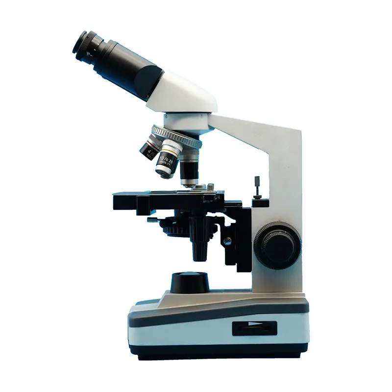
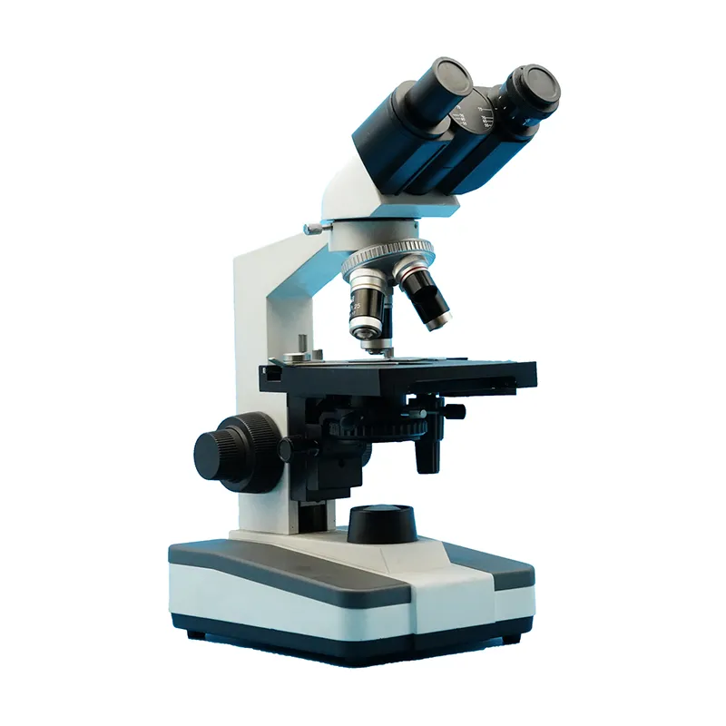
Before you start identifying blood cells, it's crucial to have a reliable binocular microscope. Our company offers a variety of high - quality microscopes, including the Binocular in Microscope, Binocular Optical Microscope, and Binocular Body Microscope. These microscopes are designed with advanced optics and ergonomic features to provide clear and comfortable viewing.
For blood cell identification, a compound binocular microscope with a high magnification power (usually 400x - 1000x) is recommended. The binocular design allows for more comfortable and accurate viewing, reducing eye strain during prolonged sessions. Our microscopes are equipped with high - quality lenses that can resolve fine details of blood cells, ensuring accurate identification.
Preparing the Blood Sample
The first step in identifying blood cells is to prepare a proper blood sample. A thin blood smear is typically used. Here's how you can prepare one:
- Clean the slides: Use lint - free tissue to clean two glass microscope slides to remove any dust or debris.
- Obtain a blood sample: This should be done following proper medical procedures. A small drop of blood is placed at one end of the clean slide.
- Make the smear: Use a second slide to spread the blood evenly over the first slide. Hold the second slide at a 30 - 45 - degree angle and gently drag it across the blood drop. The blood should spread out in a thin, even layer.
- Let the smear dry: Allow the blood smear to air - dry completely. This usually takes a few minutes.
- Fix the smear: You can fix the smear by immersing it in methanol for a few seconds. This helps to preserve the cells.
- Stain the smear: Staining is essential for better visualization of blood cells. A commonly used stain is the Wright - Giemsa stain. Follow the staining protocol carefully, which usually involves applying the stain, waiting for a specific period, and then washing it off gently.
Identifying Different Types of Blood Cells
Red Blood Cells (Erythrocytes)
Once you have your prepared and stained blood smear under the binocular microscope, start by using the low - power objective (40x) to locate the area with well - spread cells. Then, switch to the high - power objective (400x or 1000x).
Red blood cells are the most abundant cells in the blood. They appear as small, round, biconcave discs. They do not have a nucleus and have a characteristic red - orange color when stained with Wright - Giemsa stain. In a normal blood smear, red blood cells are evenly distributed, and they have a pale - staining center due to their biconcave shape.
White Blood Cells (Leukocytes)
White blood cells are less abundant than red blood cells but are larger in size. There are several types of white blood cells, and they can be further classified into two main groups: granulocytes and agranulocytes.
Granulocytes
- Neutrophils: These are the most numerous type of white blood cells. They have a multilobed nucleus (usually 3 - 5 lobes) and fine, pale - purple granules in the cytoplasm. Neutrophils are the first responders to infections and are involved in phagocytosis.
- Eosinophils: Eosinophils have a bilobed nucleus and large, bright - red granules in the cytoplasm. They are involved in the defense against parasitic infections and allergic reactions.
- Basophils: Basophils are the least common type of granulocytes. They have a large, irregularly shaped nucleus that is often obscured by large, dark - blue or purple granules in the cytoplasm. Basophils are involved in allergic responses, releasing histamine and other mediators.
Agranulocytes
- Lymphocytes: Lymphocytes can be small or large. Small lymphocytes have a large, round nucleus that nearly fills the cell, with a thin rim of light - blue cytoplasm. They are a key part of the adaptive immune system, playing roles in antibody production and cell - mediated immunity.
- Monocytes: Monocytes are the largest of the white blood cells. They have an indented or kidney - shaped nucleus and a pale - blue cytoplasm with fine granules. Monocytes can differentiate into macrophages, which are involved in phagocytosis and antigen presentation.
Platelets (Thrombocytes)
Platelets are small, irregularly shaped cell fragments. They are much smaller than red and white blood cells. Platelets often appear in clusters and have a purple - blue color when stained. Their main function is to form blood clots by aggregating at the site of injury.
Troubleshooting and Tips
- Poor image quality: If you notice a blurry or unclear image, check the lenses for dirt or debris. Clean the lenses using lens paper and a suitable lens - cleaning solution. Also, make sure the microscope is properly focused.
- Uneven staining: Uneven staining can make it difficult to identify cells accurately. This may be due to improper staining techniques. Make sure you follow the staining protocol precisely, including the staining time and washing steps.
- Cell overlapping: If the cells are overlapping, it may be because the blood smear was not made properly. Try preparing a new smear, paying more attention to spreading the blood evenly.
Conclusion
Identifying different types of blood cells under a binocular microscope is a valuable skill that can contribute to medical diagnosis and research. With the right binocular microscope, such as those available from our company (Binocular in Microscope, Binocular Optical Microscope, and Binocular Body Microscope), and following the proper preparation and identification procedures, you can achieve accurate results.
If you are interested in purchasing a high - quality binocular microscope for blood cell identification or other scientific applications, please feel free to contact us for procurement and to start a discussion about your specific needs.
References
- Jensen, G. R. (2021). "Medical Clinical Laboratory Techniques: A Pocket Guide." Pearson.
- Rodak, B. F., & Carr, J. H. (2017). "Clinical Hematology ATLAS." Elsevier.


